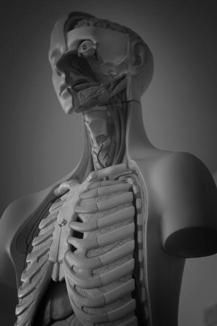
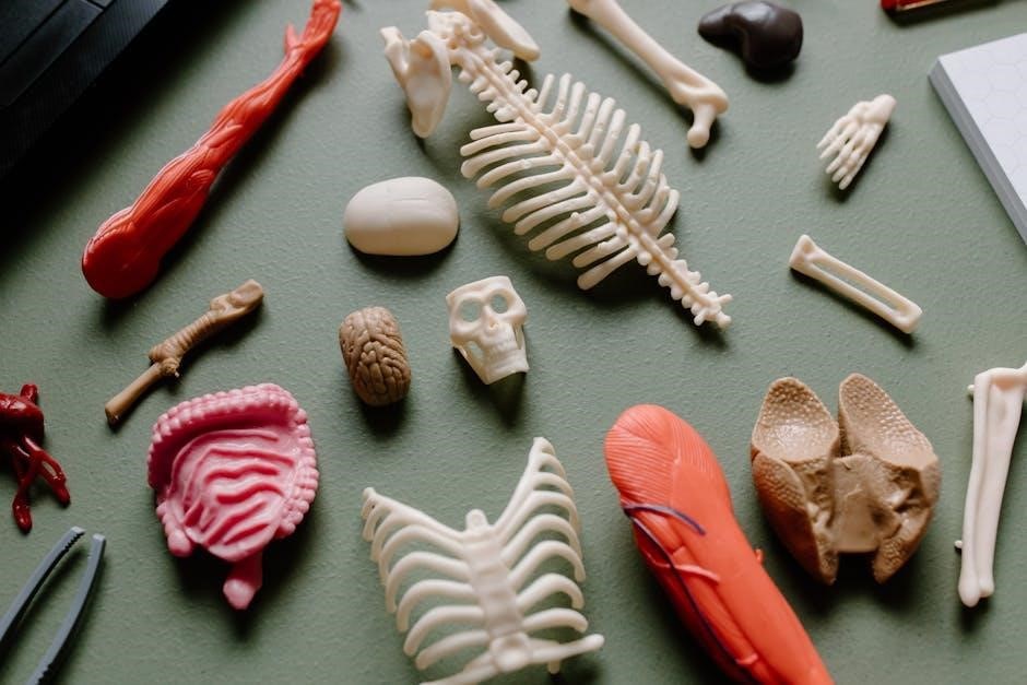
Human skeletal structure, comprised of 206 bones, provides support, protection, and enables movement․ Detailed diagrams illustrate these interconnected elements, crucial for forensic analysis and anatomical study․
Overview of the Human Skeletal System
The human skeletal system is a complex and vital framework, typically consisting of 206 bones in adulthood․ This intricate system provides structural support, protects delicate organs like the brain and heart, and facilitates movement through muscle attachment․ Initially, infants possess more bones, which fuse together during growth․
The skeleton isn’t static; it continuously remodels itself, responding to stresses and strains․ Cranial bones interlock at sutures, and growth continues for years, with facial bones developing until around age 25․ Studying skeletal elements, like those detailed in forensic literature, allows for population-specific data analysis, utilizing dimensions and sex determination via pelvic examination․ Understanding this system is fundamental to fields like anthropology and medicine․
Importance of a Diagram for Understanding Bone Structure
Visualizing the human skeleton through diagrams is paramount for comprehension․ Illustrations clearly depict the 206 bones, their intricate connections, and key anatomical features․ These diagrams aid in identifying cranial bones – frontal, parietal, temporal, occipital, sphenoid, and ethmoid – and facial structures like the mandible and maxilla;
Detailed representations showcase suture lines where bones interlock, and highlight growth patterns, including fontanelles in infants․ Diagrams are essential for forensic studies, enabling accurate measurements and sex determination․ They facilitate understanding of bone thickening with age and the eventual obliteration of sutures, providing a crucial foundation for anatomical knowledge․
The Axial Skeleton (80 Bones)
The axial skeleton, consisting of 80 bones, forms the central body structure, including the skull, vertebral column, and thoracic cage, providing vital support․
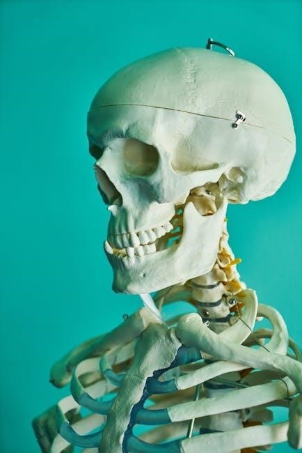
Skull (22 Bones)
The human skull, a complex structure, typically comprises 22 bones – excluding the middle ear ossicles․ It protects the brain and supports facial features․ The cranium, forming the braincase, consists of eight bones interlocked by sutures․ These include the frontal, parietal (paired), temporal (paired), occipital, sphenoid, and ethmoid․
Facial bones, totaling 14, create the face’s framework․ These encompass the nasal (paired), maxilla (paired), zygomatic (paired), mandible, lacrimal (paired), palatine (paired), inferior nasal conchae (paired), and vomer․ At birth, cranial bones aren’t fully formed, with fontanelles ossifying over the first two years of life․ Cranial vault growth continues until around 15 years, while facial skeleton growth may extend to 25․
Cranial Bones (8 Bones) ー Frontal, Parietal, Temporal, Occipital, Sphenoid, Ethmoid
The neurocranium, or braincase, is formed by eight cranial bones providing crucial protection for the brain․ The frontal bone forms the forehead, while paired parietal bones create the sides and roof of the cranium․ Temporal bones house the middle and inner ear structures․ The occipital bone forms the posterior base and foramen magnum․
The sphenoid, a complex butterfly-shaped bone, contributes to the base of the skull and orbits․ Lastly, the ethmoid bone separates the nasal cavity from the brain․ These bones interlock at sutures, fibrous joints that gradually fuse with age․ Initial development involves incomplete ossification at birth, with fontanelles closing by two years․
Facial Bones (14 Bones) ー Nasal, Maxilla, Zygomatic, Mandible, Lacrimal, Palatine, Inferior Nasal Concha, Vomer
The facial skeleton, excluding the mandible and hyoid, comprises 14 bones essential for orofacial structure and sensory support․ Paired nasal bones form the bridge of the nose, while the maxilla creates the upper jaw and contributes to the orbits․ Zygomatic bones form the cheekbones, and the mandible is the lower jaw – the only movable facial bone․
Lacrimal bones form part of the eye socket, and palatine bones contribute to the hard palate․ Inferior nasal conchae increase the surface area within the nasal cavity, and the vomer forms the inferior part of the nasal septum․ These bones work together for food processing and sensory functions․
Vertebral Column (26 Bones)
The vertebral column, a crucial component of the axial skeleton, typically consists of 26 bones providing central support and protecting the spinal cord․ It’s divided into cervical, thoracic, and lumbar regions, each with distinct characteristics․ These vertebrae articulate to allow for flexibility and movement while maintaining structural integrity․
Developmentally, the occipital bone initially comprises four parts, and the frontal bone is divided sagittally․ During the first two years of life, fontanelles – fibrous tissue membranes – ossify, completing individual cranial bones․ Growth continues until approximately 15 years, with facial skeleton growth potentially extending to 25 years․
Cervical Vertebrae (7)
The cervical vertebrae, numbering seven, form the uppermost section of the vertebral column, supporting the head and enabling its diverse range of motion․ These vertebrae are uniquely characterized by the presence of transverse foramina, facilitating the passage of vertebral arteries․ The atlas (C1) and axis (C2) are specialized, allowing for nodding and rotational movements of the head, respectively․
Cranial bone development involves ossification of fibrous tissue membranes called fontanelles during the first 24 months of life․ Growth in the neurocranium typically ceases around 15 years, while facial skeleton growth may continue until 25․
Thoracic Vertebrae (12)
The twelve thoracic vertebrae articulate with the ribs, forming the structural basis of the thoracic cage․ These vertebrae exhibit distinctive features, including costal facets for rib attachment and a generally larger size compared to cervical vertebrae․ They provide crucial support for the rib cage, protecting vital organs like the heart and lungs․
The human cranium, a large globular vessel, protects the brain and supports muscles․ Excluding the mandible and hyoid, it typically consists of 27 bones interlocking at sutures, with growth continuing until approximately 15 years․
Lumbar Vertebrae (5)
The five lumbar vertebrae are the largest and strongest of the vertebral column, designed to bear the majority of the body’s weight․ They are characterized by their robust structure and lack of costal facets for rib articulation․ These vertebrae provide stability and support for the lower back, enabling a wide range of movements․
The cranium’s bones, initially incomplete at birth with fontanelles ossifying within 24 months, interlock at sutures․ Facial skeleton growth may continue until 25 years, while cranial vault bones thicken with age․
Thoracic Cage (25 Bones)
The thoracic cage, comprising 25 bones, provides essential protection for vital organs like the heart and lungs․ It’s formed by the sternum, ribs, and thoracic vertebrae, creating a robust bony shield․ The ribs articulate with the vertebrae, enabling flexibility during respiration and movement․
The human cranium, a globular vessel protecting the brain, consists of 27 bones (excluding mandible and hyoid) interlocking at sutures․ Growth continues until approximately 15 years, with facial skeleton development potentially extending to 25 years․
Sternum (1)
The sternum, a single, flat bone, forms the anterior midline of the thoracic cage․ It provides attachment points for the ribs via cartilage, contributing to the cage’s structural integrity and flexibility during breathing․ The sternum’s development, like other cranial bones, involves ossification of fibrous tissue membranes during early life․
Cranial bones initially incomplete at birth, with fontanelles ossifying within the first 24 months․ The cranium supports masticatory muscles and sensory systems, undergoing continued thickening and suture obliteration in adulthood․
Ribs (24) ⎻ True, False, and Floating Ribs
Twenty-four ribs compose the thoracic cage, articulating with the vertebral column posteriorly and most connecting to the sternum via costal cartilage anteriorly․ These are categorized as true, false, and floating ribs․ True ribs (1-7) directly attach to the sternum, while false ribs (8-10) connect indirectly through the cartilage of ribs above․
Floating ribs (11-12) lack anterior attachment, offering less protection․ Like the cranium, rib growth continues until approximately 25 years, influenced by factors like tooth eruption and nasopharynx development․
The Appendicular Skeleton (126 Bones)
The appendicular skeleton, with 126 bones, includes limbs and girdles, enabling movement․ Studying these bones aids in understanding human locomotion and skeletal variations․
Upper Limbs (64 Bones)
The upper limbs consist of 64 bones, facilitating a wide range of movements and interactions with the environment․ This includes the shoulder girdle, arm, forearm, and hand․ The shoulder girdle, comprising the clavicle and scapula, connects the upper limb to the axial skeleton, providing flexibility․
The arm features the humerus, a single bone providing structural support․ The forearm contains the radius and ulna, enabling rotation and complex hand movements․ Finally, the hand, with its 54 bones – carpals, metacarpals, and phalanges – allows for precise manipulation and grasping; Understanding the arrangement of these bones is vital for analyzing skeletal remains and comprehending human dexterity․
Shoulder Girdle (4 Bones) ー Clavicle, Scapula
The shoulder girdle, composed of four bones (two clavicles and two scapulae), serves as the crucial link between the upper limbs and the axial skeleton․ The clavicle, or collarbone, is a slender, S-shaped bone that connects the sternum to the scapula, providing stability and transmitting forces․
The scapula, or shoulder blade, is a flat, triangular bone that articulates with the clavicle and humerus, enabling a wide range of arm movements․ This arrangement allows for significant flexibility and mobility of the upper limb, essential for various functions․ Analyzing these bones aids in determining age, sex, and stature in skeletal analysis․
Arm (2 Bones) ⎻ Humerus
The arm consists of a single bone, the humerus, extending from the shoulder to the elbow․ This long bone features distinct anatomical landmarks crucial for muscle attachment and joint articulation․ The proximal end of the humerus articulates with the scapula at the shoulder joint, enabling a wide range of motion․
Distally, it forms the elbow joint with the radius and ulna of the forearm․ The humerus’s robust structure provides stability and leverage for upper limb movements․ Forensic analysis of the humerus can reveal information about an individual’s height and potential trauma, contributing to skeletal identification․
Forearm (4 Bones) ー Radius, Ulna
The forearm comprises two bones: the radius and ulna, working in concert to enable rotation and complex movements of the hand and wrist․ The radius, located on the thumb side, allows for pronation and supination – the ability to turn the palm up and down․ The ulna, on the pinky side, provides stability to the elbow joint․
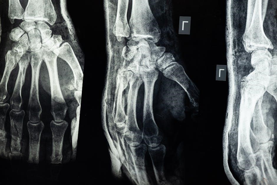
These bones articulate with the humerus at the elbow and the carpals at the wrist․ Forensic anthropologists utilize the radius and ulna’s dimensions for sex estimation and individual identification, alongside assessing potential fracture patterns․
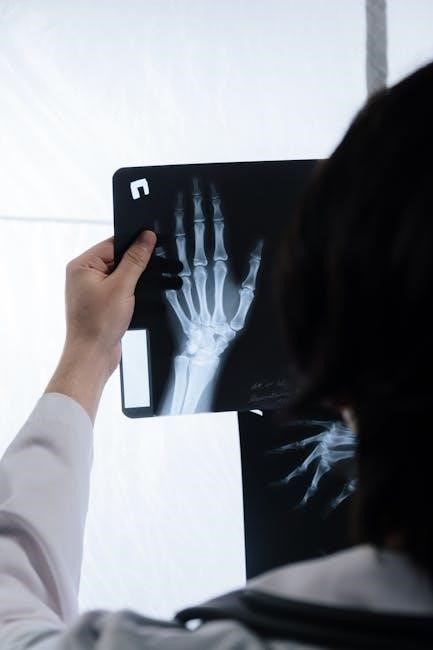
Hand (54 Bones) ー Carpals, Metacarpals, Phalanges
The human hand is a remarkably complex structure, containing 54 bones distributed across the carpals (wrist – 8 bones), metacarpals (palm – 5 bones), and phalanges (fingers – 14 bones per hand)․ These bones enable a wide range of precise movements, crucial for manipulation and interaction with the environment;
Carpal bone dimensions are utilized in forensic anthropology for sex determination and ancestry estimation․ Analyzing hand bone morphology aids in identifying individuals and understanding developmental variations․ Fracture patterns within these bones can also reveal information about trauma or activity patterns․
Lower Limbs (62 Bones)
The lower limbs, comprising 62 bones, are fundamental for weight-bearing, locomotion, and maintaining balance․ This includes the pelvic girdle (2 bones – hip bones formed by the ilium, ischium, and pubis), the femur (thigh – 2 bones), the tibia and fibula (leg – 2 bones), and the foot (52 bones – tarsals, metatarsals, and phalanges)․
Pelvic bone morphology is particularly significant in forensic anthropology for sex determination, as differences exist between male and female pelvises․ Lower limb bone length and robusticity provide insights into stature and activity patterns․ Studying these bones aids in reconstructing an individual’s lifestyle and identifying potential pathologies․
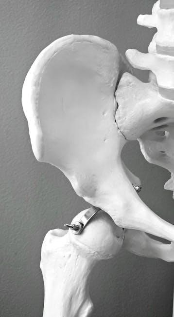
Pelvic Girdle (2 Bones) ⎻ Hip Bones (Ilium, Ischium, Pubis)
The pelvic girdle, consisting of two hip bones, connects the axial skeleton to the lower limbs, supporting weight and facilitating locomotion․ Each hip bone is formed by the fusion of three bones: the ilium, ischium, and pubis․ These bones articulate with each other and the sacrum, creating a strong and stable structure․
The pelvis exhibits significant sexual dimorphism, making it crucial for sex determination in skeletal remains․ Features like the subpubic angle and the shape of the sciatic notch differ between males and females․ Analyzing these characteristics provides valuable insights in forensic anthropological investigations․
Thigh (2 Bones) ⎻ Femur
The femur, or thigh bone, is the longest and strongest bone in the human body, extending from the hip to the knee․ It bears significant weight and plays a vital role in locomotion․ The femur’s robust structure is essential for supporting the body’s mass during activities like walking, running, and jumping․
Measurements of the femur, including its length and diameter, are frequently used in forensic anthropology to estimate stature․ Variations in femoral dimensions can also provide clues about an individual’s sex and ancestry․ Detailed analysis of the femur contributes significantly to building a biological profile from skeletal remains․
Leg (2 Bones) ー Tibia, Fibula
The lower leg consists of two bones: the tibia and the fibula․ The tibia, or shinbone, is the larger and weight-bearing bone, located medially․ It articulates with the femur at the knee and the talus at the ankle, transmitting weight from the upper leg to the foot․ The fibula, situated laterally, is a slender bone primarily involved in muscle attachment and ankle stabilization․
Forensic analysis of the tibia and fibula can reveal information about trauma, disease, and activity patterns․ Measurements of these bones contribute to stature estimation, alongside the femur․ Their robust nature often preserves valuable evidence in skeletal remains, aiding in identification and reconstruction of events․
Foot (52 Bones) ー Tarsals, Metatarsals, Phalanges
The human foot, a complex structure with 52 bones, is divided into tarsals, metatarsals, and phalanges․ The tarsals (7 bones) form the ankle and posterior part of the foot, providing stability and shock absorption․ Metatarsals (5 bones) comprise the midfoot, connecting the tarsals to the toes․ Phalanges (14 bones) form the toes, enabling propulsion and balance․
Detailed examination of foot bones aids in forensic identification, revealing information about gait, trauma, and occupation․ Variations in bone density and morphology can indicate habitual activities․ Accurate skeletal analysis of the foot is crucial for reconstructing an individual’s lifestyle and circumstances․
Understanding Bone Diagrams and Anatomical Terminology
Bone diagrams utilize specific terms to identify features and locations․ Comprehending anatomical language – sutures, processes, and foramina – is vital for accurate skeletal analysis․
Key Features to Identify on a Bone Diagram
When examining bone diagrams, several key features demand attention․ Sutures, the interlocking lines between cranial bones, are crucial for identifying skull fragments and age estimation․ Processes – bony projections – serve as muscle attachment sites, revealing information about physical activity․ Foramina, or openings, accommodate nerves and blood vessels, indicating pathways and potential injury locations․
Furthermore, observe bone thickness and texture, which change with age and sex․ Identifying landmarks like the mastoid process (temporal bone) or the mental foramen (mandible) aids in specific bone identification․ Understanding the neurocranium and orofacial skeleton distinctions is also essential․ Detailed diagrams, like those found in forensic literature, highlight these features for accurate analysis․
Common Anatomical Terms Used in Skeletal Studies
Skeletal studies rely on precise anatomical terminology․ “Neurocranium” refers to the cranial vault protecting the brain, while the “orofacial skeleton” encompasses facial bones involved in food processing․ “Fontanelles,” fibrous membranes in infants, ossify into cranial bones․ Understanding “sutures,” the interlocking joints between skull bones, is vital for age estimation․
Terms like “foramen” (opening for vessels/nerves) and “process” (bony projection for muscle attachment) are frequently encountered․ “Ossification” describes bone formation, and “obliterated” signifies suture closure with age․ Recognizing these terms, alongside descriptions of bone dimensions and variations, is crucial for accurate skeletal analysis and interpretation of forensic data․